Thyroid ultrasound images
Thyroid ultrasound is a noninvasive imaging procedure used to assess the thyroid gland for abnormalities. The thyroid gland is located in the neck and produces hormones that control metabolism. Ultrasound images of the thyroid gland can be used to evaluate the size and shape of the gland, as well as any nodules or masses that may be present.
Thyroid ultrasound images are often performed as part of a routine physical exam or if a doctor suspects a patient has a thyroid disorder. CPT code 2023 is used to report a diagnostic thyroid ultrasound. This code involves the use of real-time ultrasonography to image the thyroid gland and may also include Doppler imaging to assess blood flow within the gland.
What is Thyroid Ultrasound Images
There are several different types of thyroid ultrasound images, and each can provide valuable information about the health of your thyroid gland. A thyroid ultrasound images can help identify benign or malignant nodules, as well as assess the size and function of your thyroid gland. Thyroid ultrasound images are usually quick and painless, and can be done on an outpatient basis.
Article About:- Health & fitness
Article About:- Medical Technology
Article About:- Sports

What is Thyroid Ultrasound cpt Code
CPT code 2023 is used to report a diagnostic thyroid ultrasound. This code involves the use of real-time ultrasonography to image the thyroid gland and may also include Doppler imaging to assess blood flow within the gland.
Normal Thyroid Ultrasound
- Normal thyroid ultrasound images usually show a well-defined gland with smooth borders and no evidence of nodules, cysts, or other abnormalities. Echogenicity (hyperechoic compared to the surrounding soft tissue) is usually uniform throughout the gland.
- In some cases, small areas of calcification may be seen within the gland, but this finding is not considered abnormal. The presence of a dominant follicle is also considered normal and should not be a cause for concern.
- If no lumps or other abnormalities are identified on thyroid ultrasound, further investigation with additional imaging (eg, CT scan) or biopsy may rule out thyroid cancer or other malignancy.
What is cpt thyroid ultrasound
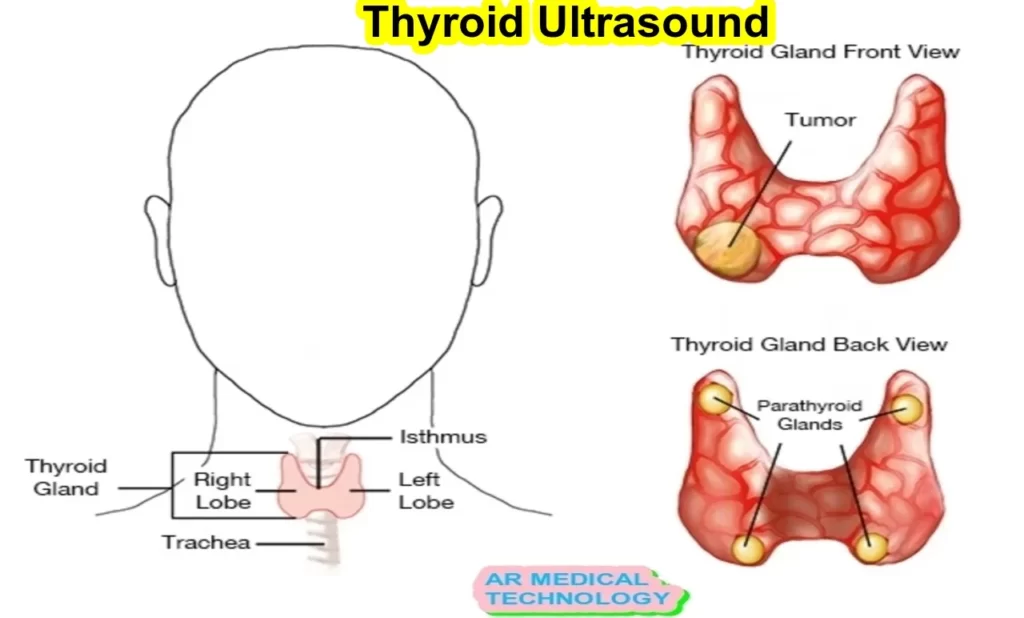
Thyroid ultrasound is used to produce images of the thyroid gland. The thyroid is a small, butterfly-shaped gland located in the front of the neck. It produces hormones that regulate the body’s metabolism.
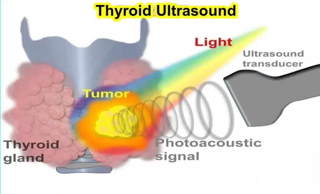
Ultrasound is a painless and non-invasive test that uses sound waves to create images of the inside of the body. During a thyroid ultrasound, a transducer (a handheld device) is placed on the skin over the thyroid gland. The transducer emits sound waves that bounce off the thyroid and are converted into images on a computer screen.
The purpose of a thyroid ultrasound is to evaluate the size, shape, and appearance of the gland. Ultrasound may also be used to locate a nodule (lump) or growth in a gland. Thyroid ultrasound is often done as part of a routine physical exam or when someone has symptoms (such as a goiter or hoarseness) that may be related to problems with the thyroid gland.
What is Thyroid Ultrasound cpt
Thyroid ultrasound is used to assess the size, shape, and texture of the thyroid gland. This procedure is done by placing a transducer (a small hand-held instrument) on the skin over the thyroid gland. The transducer emits sound waves that produce images of the thyroid gland on a monitor.

The purpose of a thyroid ultrasound is to evaluate the structure of the gland and to detect any abnormalities, such as nodules or cysts. This information can help diagnose or manage conditions such as goiter, Hashimoto’s disease, Graves’ disease, and cancer.
A thyroid ultrasound is generally safe and does not require any special preparation. However, you may be asked to avoid eating or drinking for several hours beforehand if you are also having a fine needle biopsy (a procedure in which a needle is used to collect tissue samples from the thyroid gland). ). ,
The images produced by thyroid ultrasound are typically used in conjunction with other tests, such as blood tests and imaging studies, to make a diagnosis.
Hashimoto Thyroid Ultrasound
The thyroid gland is a butterfly-shaped endocrine gland usually located in the lower part of the neck. The gland produces thyroid hormones, which are responsible for regulating metabolism.
A thyroid ultrasound is performed by placing a transducer on the skin over the thyroid gland and collecting images of the gland using sound waves. The images are then interpreted by a radiologist.
Thyroid ultrasounds are generally safe and well tolerated. There is no radiation risk associated with this test.
If you have been ordered to have a thyroid ultrasound, it is important to follow your doctor’s instructions about how to prepare for the test. You may be asked to drink water or use an iodinated contrast agent before the test to improve visualization of the gland.
MEDVICE Rechargeable Tens Unit Muscle Stimulator, 2nd Gen 16 Modes & 8 Upgraded Pads for Natural Pain Relief & Management, FDA Cleared Electric Pulse Impulse Mini Massager Machine. thyroid ultrasound images.
- PROFESSIONAL PAIN RELIEVER TENS UNIT
- UPGRADED TENS UNIT PADS
- 16 PRE-PROGRAMMED MODES/INTENSITY ADJUSTMENT
- RECHARGEABLE LITHIUM BATTERY
- ENJOY ON THE GO WITH THE PORTABLE DESIGN
FOR MORE DETAILS OF THIS PRODUCT

FAQ
What does a thyroid ultrasound show?
It is a painless, no-risk procedure for diagnosing tumors, cysts, or goiters of the thyroid gland using a hand-held ultrasound machine.
What is normal range for thyroid ultrasound?
Adult females have a thyroid volume of 10 to 15 mL and adult males have a thyroid volume of 12 to 18 mL.
What is normal size of thyroid?
In newborns, the thyroid lobe length (L or craniocaudal) measures 1.8 to 2.0 cm; in adults, it measures 4.0 to 6.0 cm; in newborns, the A-P dimension measures 0.8 to 0.9 cm; in adults, it measures 1.3 to 1.8 cm.
Why ultrasound is important for thyroid?
An equivocal physical examination confirms the presence of thyroid nodules. The purpose of this procedure is to determine whether the nodules are benign or malignant, i.e., measure their dimensions accurately and identify their internal structure and vascularization. Sonographic appearance can be used to distinguish benign thyroid masses from malignant thyroid masses.
Can ultrasound detect thyroid problems?
An ultrasound can also check an underactive or overactive thyroid gland. You may receive a thyroid ultrasound as part of an overall physical exam. Ultrasounds can provide high-resolution images of your organs that can help your doctor better understand your general health.


