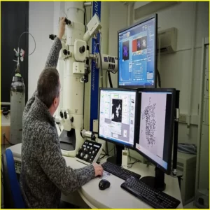
electron microscope Virus
Why is an electron microscope Virus Important?
We all know that viruses are incredibly small – too small in fact to be seen with the naked eye. So how can we see them in detail? Enter the electron microscope Virus, an advanced imaging tool that helps us see even the smallest particles In this article, find out why this incredible technique is so important to virology and what makes it so valuable to research .
What is an Electron Microscope Virus?
An electron microscope virus is a very small, spore-forming bacterium that is used to infect other cells. The viral particle, or virion, consists of a protein coat surrounding a core of genetic material. This virus can be as small as 10 nanometers in diameter. When the virus comes into contact with a suitable host cell, it attaches itself and injects its genetic material into the host cell. The viral genetic material then takes over the machinery of the host cell and causes it to produce more copies of the virus. The new viruses are then released from the host cell, where they can infect other cells.
Article About:- Health & fitness
Article About:- Medical Technology
Article About:- IR News
Article About:-Amazon Product Review

Why is an electron microscope virus used for?
One of the reasons why an electron microscope is used for viruses is that they are very efficient at replicating themselves. This means they can quickly make large numbers of copies of themselves, which can be used to infect other cells. Additionally, electron microscopes have very high resolution, which allows them to see very small details. This makes them ideal for studying viruses and other small particles. electron microscope virus
Why is it used to detect viruses?
An electron microscope is a powerful tool that can be used to detect and study viruses. Viruses are very small, and they can only be seen using an electron microscope. This type of microscope uses a beam of electrons to magnify objects.
Electron microscopes are used to study viruses because they can provide a lot of information about the structure of viruses. This information can be used to develop new ways to treat or prevent viral infections.
How does the microscope work?
Electron microscope virus/ The microscope uses a beam of electrons to produce an image of the sample. Electrons are focused by a magnetic field and passed through a thin sample on a metal grid. The electrons interact with the atoms in the sample, and the resulting pattern is magnified and projected onto a fluorescent screen, film, or CCD camera.
The magnification of the image is determined by the strength of the electron beam and the distance between the sample and the detector. The focus and contrast of the image is adjusted by different parameters such as voltage, current and the size of the objective lens.
How important is a virus in the world?
Viruses are a very important part of the world. They are responsible for many diseases and illnesses, but they are also responsible for most of the genetic diversity in the world. Viruses can be helpful or harmful, depending on the context in which they are found.
In the natural world, viruses play a major role in evolution by providing new genetic material to species. Viruses can also harm ecosystems, including coral reefs and forests, by killing large populations of organisms. In the medical world, viruses are used for the research and development of vaccines and other treatments for diseases.
How common are viruses?
Viruses are extremely common and can be found in almost every corner of the world. It is estimated that there are over 10 million different types of viruses in the world, with new ones being discovered all the time. Most viruses are harmless to humans and do nothing more than cause a cold or flu-like illness. However, some viruses can be quite dangerous and even fatal.


