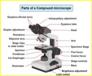
compound microscope
Compound Microscope What Does It Do?
Have you ever wondered what happens inside the laboratory? One of the most important tools for scientists is the compound microscope. In this article, learn about what a compound microscope is, how it works, and its uses in research.
What is a Compound Microscope?
A compound microscope uses two lenses to magnify an object. The first lens, the objective lens, is positioned near the specimen. The second lens, the ocular lens, is located in the eyepiece. The ocular lens magnifies the image formed by the objective lens.
Compound microscopes are used to view small objects in great detail. They are commonly used in scientific laboratories and classrooms.
Article About:- Health & fitness
Article About:- Medical Technology
Article About:- IR News
Article About:-Amazon Product Review

How Does The Microscope Work?
A compound microscope is an optical microscope that uses a combination of lenses to magnify objects. The first lens, called the objective lens, is positioned near the object being viewed. A second lens, called the eyepiece, is positioned near the observer’s eye.
The objective lens forms a real image of the object being viewed which is larger than the actual object. The eyepiece magnifies this real image so that it appears larger to the observer.
Compound microscopes can magnify objects up to 1000 times their actual size.
How to Read a Microscope Slide
To read a microscope slide, first make sure the eyepiece is clean and the light source is on. Adjust the focus knob until the image is clear. To examine different areas of the slide, move the slide around on the stage. Once you have finished examining the slide, turn off the light source and return the slide to its case.
What Do You Look For in a MicroScope Slide?
When looking for microscope slides, you want to find one that is made of high quality materials and designed for durability. You also want to look for a slide that has a smooth surface so that your specimens will not be damaged while you are viewing them. Additionally, you want to find a slide that is transparent so that you can clearly see your samples.
How to Draw A Skeleton of a Fish
Let’s say you want a step-by-step guide on how to draw a fish skeleton:
1. Begin by sketching the basic shape of the fish. If you need help, look at the picture of the fish for reference.
2. Once you have the basic shape down, start adding the main bones of the skeleton. The backbone should be the longest, and then you can add ribs and other bones as needed.
3. To finish, add small details like teeth or scales. Erase any guidelines from your initial sketch and color in, if desired.
Types of compound microscope
A compound microscope is a type of optical microscope that uses two or more lenses to magnify objects. The first lens, the objective lens, is located near the object being viewed and the second lens, the ocular lens, is located near the observer’s eye. The magnifying power of a compound microscope is determined by the ratio of the focal lengths of these two lenses.
Most compound microscopes have three or four different objective lenses that can be used to view objects at different magnifications. The highest magnifying power is usually obtained with objective lenses of shortest focal length. Compound microscopes also have an adjustable diaphragm that controls the amount of light entering the microscope, as well as an iris that adjusts the diameter of the beam of light passing through the object being viewed.
There are several different types of compound microscopes, including those with monocular heads, binocular heads, trinocular heads, and those with inverted optics. Monocular microscopes have one eyepiece, while binocular and trinocular microscopes have two and three eyepieces, respectively. Inverted microscopes have their optics inverted so that the specimen is viewed from below rather than from above.


