normal brain mri vs ms, normal brain mri vs ms
Normal Brain MRI Vs MS
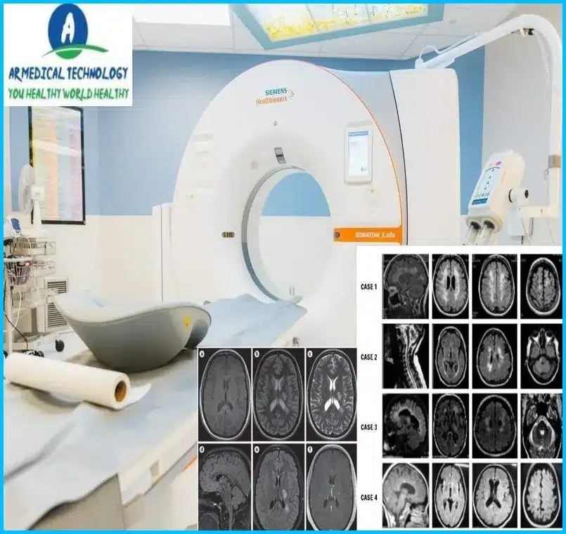
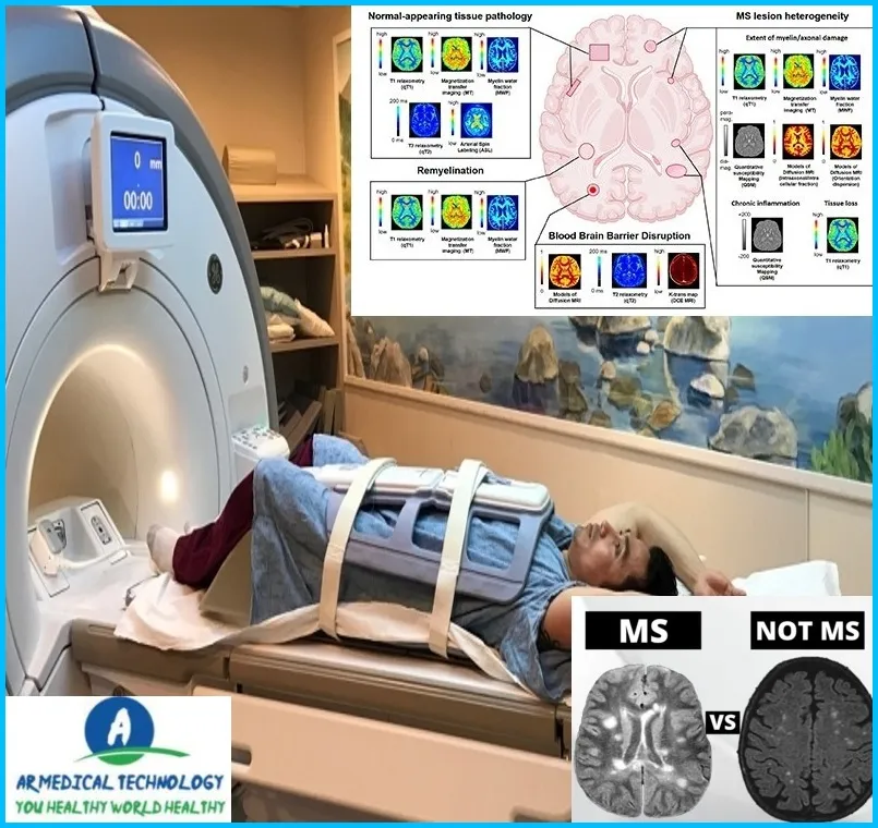
An aberrant brain MRI of a young MS patient is seen here in comparison to a normal brain MRI. Several lesions may be seen in this scan as bright white dots. A person without multiple sclerosis would have a more evenly grayed brain with fewer bright spots on a brain scan.
Welcome to our blog article on MS vs. Normal Brain MRI! Determining exactly what’s going through your head when faced with a probable multiple sclerosis diagnosis might be one of the most irritating things. Given that the symptoms could vary from little tingling and numbness to complete immobility, it seems sense to seek treatment as soon as feasible.
It’s crucial to recognize the differences between an MRI of the brain that is normal and one that exhibits symptoms of multiple sclerosis. It can assist you in developing a deeper understanding of your illness and enabling you to make wise decisions regarding your future medical care. able to. Alright, let’s examine these two MRI kinds side by side.
What do MS Lesions Look like on MRI
It can be challenging to distinguish MS lesions on an MRI since they can show in a variety of ways. Nonetheless, MS lesions share a few characteristics. When seen on T2-weighted or FLAIR imaging, they often take the form of round or oval-shaped regions with elevated signal intensity. On T1-weighted images, lesions are also visible, however they often show up as darker patches. Contrast agent may occasionally be used to highlight the lesions.
Article About:- Health & fitness
Article About:- Medical Technology
Article About:- Sports

MS brain MRI vs normal
There are many different types of brain MRI, but one of the most important is the MS brain MRI. This type of MRI can help diagnose and track the progress of multiple sclerosis.
MS brain MRI differs from regular brain MRI in several ways. First, an MS brain MRI typically includes more images than a regular brain MRI. This is because MS can affect many different parts of the brain, and it is important to get detailed information about all affected areas.
Second, an MS brain MRI often uses special techniques, such as gadolinium-enhanced imaging, that are not used in regular brain MRIs. These techniques may help to better identify lesions associated with MS.
Finally, an MS brain MRI may be interpreted differently from a regular brain MRI. For example, some features that may be considered normal on a regular brain MRI may be considered abnormal on an MS brain MRI. This is because a lot is still not known about MS, and how it affects the brain.

Does early MS show up on MRI
Multiple sclerosis (MS) is a progressive neurological condition that can cause a wide range of symptoms, including problems with vision, arm or leg movement, sensation, or balance. Diagnosing early MS can be difficult because symptoms may be mild and may come and go, or they may mimic other conditions.
MRI is the best tool for diagnosing MS. An MRI scan can show lesions (areas of damage) in the brain and spinal cord that are typical of MS. The lesions appear as bright spots on MRI images.
If you have signs and symptoms that suggest you may have MS, your doctor may order an MRI scan. If an MRI scan shows lesions consistent with MS, your doctor will make a diagnosis of MS.
Does MS show up on MRI without contrast
No, MS does not show up on MRI without contrast. Contrast is needed to see the lesions associated with MS.
Normal vs MS brain MRI images
There are many different types of brain MRI images, but the two most common are normal brain MRI images and MS brain MRI images. Both types of images can be used to diagnose a variety of conditions, but there are some important differences between them.
Normal brain MRI images are taken without contrast, meaning they do not show any lesions or other abnormalities. MS brain MRI images, on the other hand, are taken with contrast, which allows doctors to see lesions and other abnormalities. The main difference between the two types of images is that normal brain MRI images do not show any evidence of disease, whereas MS brain MRI images do.
If you’re unsure what type of image you need, your doctor can help you decide. In general, however, normal brain MRI images are best for diagnosing conditions that do not involve the central nervous system, whereas MS brain MRI images are best for diagnosing conditions that involve the central nervous system.

FAQ
How does a MRI show MS vs normal?
The fat is removed from the regions where MS has destroyed the myelin. Once the fat is gone, the region becomes more water-filled and, depending on the type of scan, appears as either a dazzling white spot or a darkish area on an MRI.
Will a brain MRI show signs of MS?
an MRI. The most typical course of action is to undergo an MRI (magnetic resonance imaging) of your brain and/or spinal cord. The scars left by MS, known as lesions, are visible as tiny white spots and may be found with this scan.
Does white spots on brain mean MS?
White patches on an MRI can occur in conditions other than multiple sclerosis, though. “According to a study that we published in 2019, nearly 1 in 5 new patients coming into our clinic with an existing diagnosis of MS turned out not to actually have MS,” Kaisey stated.
Can I have MS if my MRI is normal?
A normal brain MRI does not automatically rule out the potential of multiple sclerosis, despite MRI being a highly helpful diagnostic technique. Approximately 5% of individuals with MS who receive a diagnosis do not initially exhibit brain lesions as seen by an MRI.
What are usually the first signs of MS?
Ailments
Typically, one side of the body has numbness or paralysis in one or more limbs at a time.
prickling.
Sensations of electric shock brought on by specific neck motions, particularly bending the neck forward (Lhermitte sign)
Insufficient coordination.
shaky walking or unsteadiness in stride.


