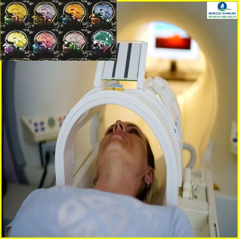
brain mri with contrast
How To Interpret Brain Mri With Contrast
One of the most crucial diagnostic methods for neurological disorders is a brain MRI. But how are these really interpreted by doctors? We will look at how brain MRIs operate and how medical professionals utilize them to identify neurological disorders in this blog article. We will also discuss some of the many forms of contrast used in brain MRIs and how they might improve the image that medical professionals see of the brain’s internal workings.
Brain Mri With Contrast
Many details on the composition and operation of the brain may be obtained from a brain MRI with contrast. The contrast agent aids in the highlight of certain brain regions, which is useful in the diagnosis of diseases including Alzheimer’s, stroke, and malignancies.
When analyzing a brain MRI with contrast, it’s critical to examine the entire image rather than simply concentrating on one spot. While certain locations may seem brighter than others due to the contrast agent, this does not always indicate that there is an abnormality in that area.
Brain MRIs with contrast come in a variety of forms, and each has advantages and disadvantages of its own. It is crucial that you collaborate with a licensed radiologist who can support you.
Article About:- Health & fitness
Article About:- Medical Technology
Article About:- Sports

Brain Mri With Contrast vs Without Contrast
A brain MRI with contrast can provide more detailed information about the structures of the brain than a non-contrast MRI. Contrast agents, such as gadolinium, are injected into the body before the MRI to help improve the image quality.
Brain MRIs with contrast can be used to better visualize abnormalities, such as tumors or lesions. They can also be helpful in assessing damage from strokes or other conditions that cause changes in the brain tissue.
cpt Code for Brain Mri With Contrast
The cpt code for brain mri with contrast is 78452. This code is used to indicate that a radiologist has performed a brain MRI with the help of a contrast agent. The use of contrast agents can help to improve the quality of the MRI images, and can also help to provide more information about the structure and function of the brain.
Brain Mri With Contrast cpt Code
If you’re looking for a brain MRI with contrast cpt code, you’ve come to the right place. In this article, we’ll show you how to interpret brain MRIs with contrast so that you can get the most accurate results possible.
First, it’s important to understand what contrast is and how it works. Contrast is used in medical imaging to help doctors better see certain structures or abnormalities. It’s injected into the body and then travels through the bloodstream. When it reaches the area being imaged, it helps to make that area more visible on the scan.
There are two types of contrast agents that are commonly used for brain MRIs: gadolinium-based contrast agents (GBCAs) and iodine-based contrast agents (IBCAs). GBCAs are typically used for patients who have had prior strokes or who are at risk for stroke. IBCAs are typically used for patients who have tumors or other abnormalities in their brains.
Once the contrast agent has been injected, the MRI scan will be performed. The resulting images will show areas of enhanced signal intensity, which indicate where the contrast agent has accumulated. These areas can then be further evaluated by a radiologist to look for specific features or abnormalities.
The brain MRI with contrast cpt code is 70551. This code is used when billing for an MRI of the brain that includes gadolinium-based contrast agent administration.
Abnormal Brain Mri with Contrast
An MRI with contrast uses a special dye to help highlight certain areas of the brain. This can be helpful in diagnosing conditions such as tumors, strokes, and other abnormalities.
When interpreting an MRI with contrast, it is important to look for any areas of the brain that appear darker or brighter than usual. These areas may indicate where there is an issue.
It is also important to look at the overall size and shape of the brain. Any abnormalities in this area may also be indicative of a problem.
If you are unsure of what you are seeing on an MRI with contrast, it is important to consult with a medical professional who can give you more information about what the scan is showing.

FAQ
What does a brain MRI with contrast show?

Mind. An MRI with contrast provides a more detailed image of your brain damage than one without it if you have had an accident and sustained one. In addition, it can be used to diagnose dementia, multiple sclerosis, stroke, brain infections, and malignancies of the brain.
What is the difference between a brain MRI and a brain MRI with contrast?

Important lessons learned: MRIs with and without contrast are painless and non-invasive procedures. While non-contrast MRIs do not employ contrast dyes like gadolinium or iodine, contrast MRIs do. Patients without a history of pregnancy or pre-existing medical issues, such as renal abnormalities, can safely have a contrast MRI scan.
How long does a brain MRI with contrast take?

Your doctor’s orders or the specific body portion of interest will determine how long your exam will last. Exams in general will last 45–60 minutes, whereas exams in specialization may go up to two hours. Exams for the spine and brain may take 45 minutes on average. Studying might take up to fifteen minutes longer if the test involves contrast.
Do all brain MRI need contrast?

A contrast MRI is not necessary for the majority of acute occurrences, such as acute headaches, acute cerebrovascular accidents (strokes) or transient ischemic attacks, hemorrhages, and concussions.
What are side effects of MRI with contrast?

The most common side effects are headaches, dizziness, rash, itching, nausea, and pain at the injection site. Nephrogenic systemic fibrosis 1 and gadolinium poisoning are two dangerous but infrequent adverse effects that are more common in patients with considerable renal problems.
Which MRI is best for brain?

An MRI of the head and a brain are the same process. They both offer views of your brain’s inside. While head and brain MRIs are primarily used by medical professionals to evaluate brain function, these imaging techniques may also produce pictures of other head components, including blood vessels, nerves, and facial bones.


