What Is a Liver Ultrasound
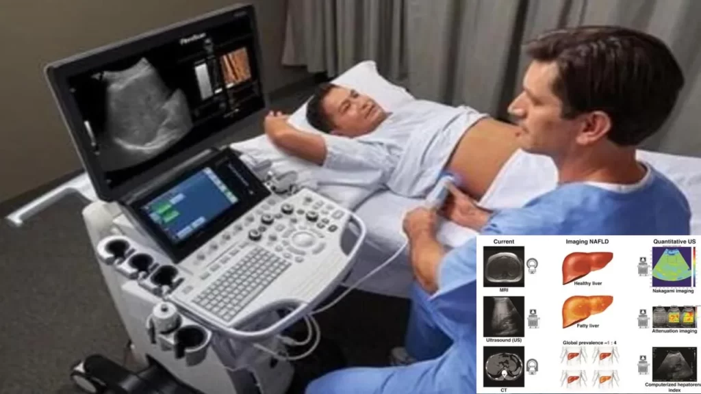
A hepatic ultrasound is a type of medical imaging that produces finely detailed pictures of the liver and the tissues around it in the abdomen by using sound waves. It is a widely used, non-invasive diagnostic technique for determining the size, form, and health of the liver. An outline of the goal, process, and uses of a liver ultrasound is provided below:
Purpose:
- Liver Structure and Size: A liver ultrasound provides information about the size, shape, and structure of the liver. Abnormalities such as enlargement, shrinkage, or changes in liver texture can be detected.
- Liver Blood Flow: The procedure allows for the assessment of blood flow within the liver’s blood vessels. Doppler ultrasound, a specific technique, can be used to evaluate the speed and direction of blood flow.
- Detection of Liver Lesions: Liver ultrasounds can help identify various liver conditions, including:
- Liver Cirrhosis: Scarring of the liver tissue.
- Hepatic Cysts: Fluid-filled sacs in the liver.
- Hepatic Hemangiomas: Benign tumors made up of blood vessels.
- Focal Liver Lesions: Such as tumors or abscesses.
- Evaluation of the Biliary System: The ultrasound can assess the bile ducts and gallbladder for any blockages, stones, or abnormalities.
- Guidance for Liver Biopsy: In some cases, a liver ultrasound may be used to guide a needle during a liver biopsy, allowing for the collection of a tissue sample for further analysis.
Procedure:
- Preparation: In most cases, little to no special preparation is required. The patient may be asked to fast for a few hours before the procedure to ensure a clear view of the liver.
- Positioning: The patient lies on an examination table, and a water-based gel is applied to the abdomen. The gel helps transmit sound waves and improve the quality of the images.
- Ultrasound Transducer: A handheld device called a transducer is moved over the abdominal area. The transducer emits high-frequency sound waves that bounce off the liver and other structures, and the echoes are converted into images on a monitor.
- Image Interpretation: A healthcare professional interprets the ultrasound images in real-time and assesses the liver’s size, texture, blood flow, and the presence of any abnormalities.
- Duration: The procedure is typically quick, taking about 15 to 30 minutes.
Post-Procedure: After the liver ultrasound, the patient can usually resume normal activities. The healthcare provider reviews the images and discusses the findings with the patient.
Liver ultrasound is a valuable tool in the diagnosis and monitoring of various liver conditions. If you have symptoms or concerns related to liver health, consult with a healthcare provider who can determine if a liver ultrasound or other diagnostic tests are necessary for a thorough evaluation.

Liver Ultrasound
Located on the upper right side of the belly, the liver is a large organ that is essential to metabolism and detoxification. High-frequency sound waves are applied to the skin covering the liver using an ultrasonic probe to produce pictures of the organ.
Though most liver ultrasonography procedures are safe and painless, there are a few things you should know before you attend. Before the exam, you must fast for at least four hours. That implies you will just consume water to eat and drink. Additionally, since oil and lotion may obstruct the ultrasonic frequencies, you should refrain from applying them to your skin. Finally, make sure the garments are easy to put on and fit comfortably around your tummy.
Article About:- Health & fitness
Article About:- Medical Technology
Article About:- Sports

Liver Ultrasound Images
There are several forms of liver ultrasonography, but the most common one is hepatic ultrasonography. This kind of ultrasonography creates a picture of your liver by using sound waves. Next, any growths or tumors that could be harming the liver are detected using the picture.
You need to fast for at least six hours before to the exam in order to be ready for a hepatic ultrasonography. In order to maintain a clean urinal, you’ll also need to drink a lot of water. It is possible that you may be asked to take some contrast material, which will improve the image of your liver during the scan.
You will be required to lie on your back on an exam table in order to get the ultrasound. The technician will apply a gel to your abdomen and capture photographs using a transducer, which resembles a wand. Sound waves are sent into your liver through the gel by the transducer. Your liver reflects these waves, which are then detected by the transducer and transformed into electrical impulses. After that, the impulses are transmitted to a computer, which produces pictures that your physician will examine.
Usually, the entire procedure takes no more than 20 to 30 minutes. After the test, there is no pain and you may resume your regular activities.

Liver Ultrasound Pre
The liver is a large organ located in the upper right side of the abdomen, just below the rib cage. The purpose of liver ultrasound is to evaluate the size, shape, and blood flow of the liver. It can also be used to detect abnormalities such as tumors or cysts.
Liver ultrasounds are usually performed as outpatient procedures. This means you will not need to stay in hospital overnight.
Prior to the liver ultrasound, you will need to fast for at least eight hours. Water is the only food or drink that should be consumed during this time. A request to refrain from taking specific medications before to the test may also be made.
You will receive detailed instructions from your doctor on how to get ready for your liver ultrasound. Make sure you properly follow these directions.
Liver ultrasonography is risk-free and typically safe. However, there is a small chance of problems like bleeding or infection, just like with any medical operation.
Liver Ultrasound cpt Code
It is used to evaluate the size, shape and blood flow of the liver. The test is also used to detect tumors, cysts, or other abnormalities.
Preparing for a liver ultrasound usually requires fasting for 8 hours prior to the procedure. You may also be asked to drink a contrast agent which will help improve the images of your liver. Once you arrive for your procedure, you will be asked to lie on your back on an exam table. A clear gel will be applied to your abdomen and a transducer will be placed on your skin. The transducer emits sound waves that are reflected from your organs and translated into images on a computer screen. The exam is usually painless and takes about 20-30 minutes to complete.

Fasting Liver Ultrasound
It has many functions, including filtering toxins from the blood and producing bile to help digest fats.
Ultrasound is a safe and painless test that can be used to examine the liver. A trained ultrasound technician will place a transducer (hand-held device) on the skin over the liver.
The transducer emits sound waves that strike the organs and tissues inside the body. These waves are converted into images that can be viewed on a monitor.
Liver ultrasound is usually performed as part of a routine physical exam or to evaluate symptoms such as abdominal pain or abnormal liver tests. They may also be ordered if someone has a history of liver disease, such as hepatitis or cirrhosis.

FAQ
What type of ultrasound is a liver ultrasound?

A liver ultrasound is a noninvasive test that produces images of the liver and its blood vessels. It can diagnose various liver conditions, such as fatty liver, liver cancer, and gallstones. It is a type of transabdominal ultrasound.
What is the special ultrasound for the liver?

As well as ultrasound elastography, transient elastography is also known. An ultrasound machine uses sound waves to send vibrations into your liver. It measures how fast the vibrations travel through your liver. Vibrations will move faster through stiff liver tissue.
What are the different types of liver scans?

The liver, gallbladder, and biliary tract can be imaged using ultrasound, radionuclide scanning, computed tomography (CT), magnetic resonance imaging (MRI), endoscopic retrograde cholangiopancreatography (ERCP), transhepatic cholangiography, and operative cholangiography.
Can ultrasound detect fatty liver?

The sensitivity and specificity of ultrasonography in detecting moderate to severe fatty liver are comparable to those of histology.
What kind of ultrasound is done for fatty liver?

The primary imaging method for patients with suspected NAFLD is transabdominal ultrasound [3]. Steatosis is characterized by an increased echogenicity of the liver parenchyma compared to the right kidney cortex, a result of intracellular fat vacuoles reflecting the ultrasound beam.
Can ultrasound detect liver damage?

Testing a tissue sample. Removing a tissue sample (biopsy) from your liver may help diagnose liver disease and look for signs of liver damage. Ultrasound, CT scan, and MRI can all show liver damage.
What are the 4 warning signs of a damaged liver?

Symptoms such as jaundice or yellowing of the eyes or skin. Pain and distention of the abdomen as a result of liver fluid release. Swelling of the lower legs as a result of fluid retention.
Confusion or forgetfulness.
Dark-colored urine.
Pale-colored stool.
Can I drink water before a liver ultrasound?
Consuming water influences the hemodynamics and stiffness of the liver. It is advised to avoid drinking any water at least an hour before liver ultrasonography elastography and Doppler sonography.


