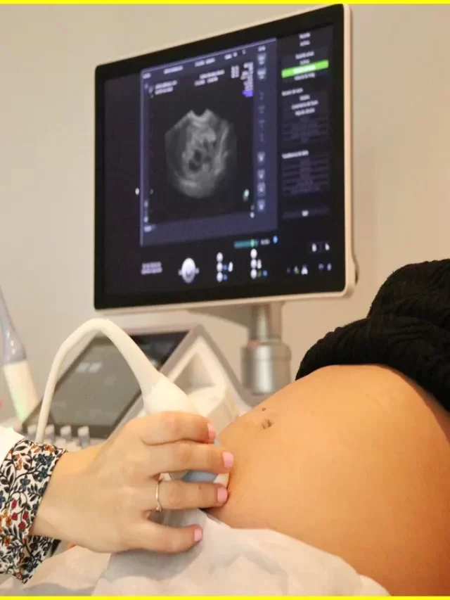
What To Expect Vaginal Ultrasound
A vaginal ultrasound is a diagnostic test that uses high-frequency sound waves to produce images of the inside of the vagina and pelvic organs. The test is also called a pelvic ultrasound or transvaginal ultrasound. A vaginal ultrasound can be used to: – Check for abnormalities in the vagina or pelvic organs – Evaluate the cause of symptoms such as pain, abnormal bleeding, or discharge – Guide procedures such as biopsies or surgery The test is usually performed by a gynecologist or radiologist, and it usually takes less than 30 minutes.
Vaginal Ultrasound
A vaginal ultrasound is a diagnostic test that uses high-frequency sound waves to produce images of the inside of the vagina and nearby structures. The procedure is also sometimes called pelvic ultrasonography or pelvic sonography.
During a vaginal ultrasound, a small transducer (probe) is inserted into the vagina. The transducer emits sound waves that bounce off organs and tissues in the pelvis to create an image (sonogram). A vaginal ultrasound can be used to:
Evaluate the size, shape, and position of the uterus
Assess the thickness of the endometrium (lining of the uterus)
Determine whether the ovaries are present and if they contain fluid-filled sacs (follicles) that may contain eggs
Detect fibroids or other abnormalities in the uterus or ovaries
The test is usually performed by a specially trained technician or doctor called a sonographer, who will interpret the images. A vaginal ultrasound is generally safe and well tolerated. There is no radiation exposure associated with this test. You may experience some discomfort from lying on your back for an extended period of time or from having something inserted into your vagina.
Article About:- Health & fitness
Article About:- Medical Technology
Article About:- IR News
Article About:-Amazon Product Review
Vaginal Ultrasound Tool
A vaginal ultrasound is a diagnostic tool used to visualize the pelvic organs and structures. The procedure is performed by inserting a small, hand-held device called a transducer into the vagina. The transducer emits sound waves that create an image of the pelvic organs on a monitor.
A vaginal ultrasound can be used to evaluate the health of the uterus, ovaries, fallopian tubes, and other structures in the pelvis. The procedure can also be used to diagnose conditions such as uterine fibroids, ovarian cysts, endometriosis, and pelvic inflammatory disease.
The vaginal ultrasound is a safe and painless procedure. There is no risk of radiation exposure with this type of ultrasound. The procedure takes about 30 minutes to complete.
Vaginal Ultrasound Pregnancy
If you’re pregnant and have had no complications, you’ll likely have at least one vaginel ultrasound during your pregnancy. A vaginal ultrasound is a diagnostic tool doctors use to visualize certain features of the fetus and uterus during pregnancy.
During a vaginel ultrasound, a small probe is inserted into the vagina. The probe emits sound waves that create an image of the fetus and uterus on a monitor.
Vaginel ultrasounds are typically performed during the second trimester of pregnancy, around 18 to 20 weeks gestation. But they may be done earlier in pregnancy if there are concerns about the health of the fetus or if the due date is uncertain.
A vaginel ultrasound can help your doctor:
* Confirm the presence of a heartbeat
* Determine how many fetuses are present
* Assess the position of the fetus
* Measure the size and development of the fetus
* Check for any birth defects or abnormalities
* Evaluate your pelvic organs
* Determine whether you have twins, triplets, or other multiples
You may be asked to empty your bladder before having a vaginal ultrasound so that your bladder doesn’t get in the way of getting clear images.
What is a Vaginal Ultrasound
A vaginal ultrasound is a diagnostic procedure that uses high-frequency sound waves to produce images of the inside of the vagina and other pelvic structures. The images may be used to help diagnose and treat conditions such as endometriosis, uterine fibroids, or pelvic inflammatory disease.
The vaginal ultrasound is usually performed by a gynecologist or other trained health care provider. The procedure is typically done in an outpatient setting, meaning you can go home the same day.
During a vaginal ultrasound, a small wand-like device (transducer) is inserted into the vagina. The transducer emits sound waves that bounce off organs and create echoes. These echoes are converted into electrical impulses that produce images on a computer screen.
You may be asked to empty your bladder before the procedure so that your bladder does not obscure the view of your pelvic organs. You may also be asked to undress from the waist down and put on a gown.
You will be positioned lying on your back on an exam table with your feet in stirrups. This allows your health care provider to have easy access to your vagina for insertion of the transducer. A gel will be applied to the transducer and then inserted into your vagina. The gel helps maintain contact between the transducer and your skin.
The transducer emits sound waves that bounce off organs and create echoes. These echoes are converted into electrical impulses that produce images on a computer.
Trans Vaginal Ultrasound
1. Transvaginal ultrasound:
A transvaginal ultrasound is an ultrasound that is performed through the vagina. This type of ultrasound can be used to assess the pelvic organs, such as the uterus, ovaries, and fallopian tubes. It can also be used to look for signs of endometriosis, fibroids, or other conditions.
A transvaginal ultrasound is generally safe and does not have any major side effects. However, you may experience some mild discomfort during the procedure. The ultrasound probe will be inserted into your vagina, and you may feel pressure or a fullness in your pelvis.
You should not eat or drink anything for at least six hours before your transvaginal ultrasound so that your bladder is full. This will help the doctor get a better view of your pelvic organs. You may also be asked to empty your bladder just before the procedure begins.
Farmscan L60 Compact Handhold Veterinary Ultrasound Scanner for Bovine, Equine, Swine pregancy, gynecological diagnosic
- Warranty: Main unit: 2 Years; Probes: 1 Year. Free Lifetime upgrade to new versions.
- Image mode: B,M, 2B, B/M, 4B; Premium image quality, considerate design for comfortable, fast and reliable detection of pregancy and gynecological diagnosic in difficlult field conditions on a dairly basis.
- Versatile and easy use for all veterinary applications on bovine, equine, swine, etc;
- Li-ion battery with 3 operating hours; Images and videos export via USB 2.0 with 450 frame cine loop inside
- Software & Report for reproductive system, and measurement for distance, area, circumference, volume, angel, heart rate, etc.


